
Comprehensive Ultrasound Guides | Expert Insights & Techniques
Discover expert insights and practical techniques with our comprehensive Ultrasound Guides. Learn how to optimize imaging and enhance diagnostic accuracy today.
Ultrasound technology has revolutionized the medical field, offering a non-invasive, safe, and effective way to diagnose and treat various conditions. For expecting mothers and those planning to conceive, ultrasound imaging plays a crucial role in ensuring the health and well-being of both the mother and the baby. This comprehensive guide will walk you through everything you need to know about ultrasound imaging, its applications, techniques, and safety measures. Whether you’re preparing for your first ultrasound or simply curious about the process, this guide is here to provide clarity and peace of mind.
What is Ultrasound Imaging?
Ultrasound imaging, also known as sonography, uses high-frequency sound waves to create real-time images of the inside of the body. Unlike X-rays, ultrasound does not use radiation, making it a safer option, especially for pregnant women. It is widely used in obstetrics to monitor fetal development, but it also has applications in diagnosing conditions in organs, muscles, tendons, and blood vessels.
Key Applications of Ultrasound in Pregnancy
Fetal Development Monitoring: Ultrasound helps track the growth and development of the baby, ensuring everything is progressing as expected.
Detecting Abnormalities: It can identify potential issues such as congenital disabilities or growth restrictions.
Placenta and Amniotic Fluid Assessment: Ultrasound checks the placenta’s position and the amount of amniotic fluid, both critical for a healthy pregnancy.
Multiple Pregnancies: It confirms the presence of twins or triplets and monitors their development.
Guiding Procedures: Ultrasound is used to guide procedures like amniocentesis or chorionic villus sampling (CVS) safely.
Types of Ultrasound Techniques
Transabdominal Ultrasound: The most common type, where a transducer is moved over the abdomen to capture images.
Transvaginal Ultrasound: Provides clearer images in early pregnancy by inserting a probe into the vagina.
Doppler Ultrasound: Measures blood flow in the baby’s heart and umbilical cord, ensuring proper circulation.
3D and 4D Ultrasound: Creates detailed, three-dimensional images and even real-time videos of the baby.
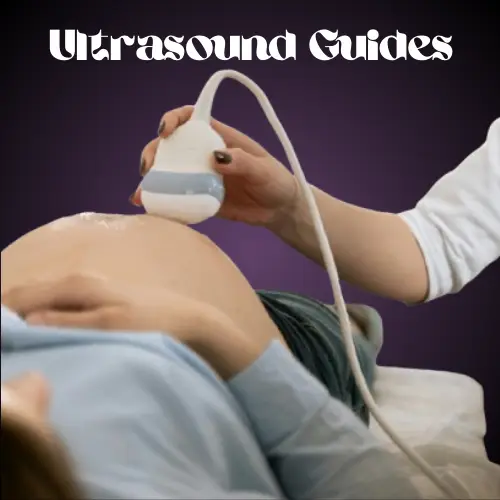
How Ultrasound Works
During an ultrasound, a trained technician (sonographer) applies a gel to the skin and uses a handheld device called a transducer. The transducer emits sound waves that bounce off tissues and organs, creating echoes. These echoes are then converted into images displayed on a monitor. The process is painless and typically takes 20-30 minutes.
Benefits of Ultrasound Imaging
Non-Invasive and Safe: No radiation is involved, making it safe for both mother and baby.
Real-Time Imaging: Provides immediate results, allowing doctors to make quick decisions.
Versatile: Used for diagnosis, monitoring, and guiding medical procedures.
Painless and Comfortable: Most women find the procedure comfortable and stress-free.
Ultrasound Safety: What You Need to Know
Ultrasound is considered safe when performed by trained professionals. However, it’s essential to follow these guidelines:
Limit Unnecessary Scans: Only undergo ultrasounds when medically recommended.
Choose Qualified Providers: Ensure the procedure is performed by certified sonographers.
Avoid Non-Medical Ultrasounds: While 3D/4D keepsake ultrasounds are popular, they should not replace medical scans.
Preparing for Your Ultrasound
Drink Water: For transabdominal ultrasounds, a full bladder helps produce clearer images.
Wear Comfortable Clothing: Loose-fitting clothes make the process easier.
Follow Instructions: Your healthcare provider will give specific guidelines based on the type of ultrasound.
FAQs About Ultrasound Imaging
1. Is ultrasound safe during pregnancy?
Yes, ultrasound is considered safe for both the mother and the baby. It uses sound waves instead of radiation, making it a preferred imaging method during pregnancy.
2. How many ultrasounds are needed during pregnancy?
Typically, two ultrasounds are recommended: one in the first trimester to confirm the due date and another in the second trimester (around 18-22 weeks) to check the baby’s development. Additional scans may be needed if there are complications.
3. Can ultrasound detect all birth defects?
While ultrasound can identify many abnormalities, it cannot detect all birth defects. Some conditions may only be diagnosed after birth.
4. What should I do if my ultrasound shows a problem?
If an issue is detected, your healthcare provider will discuss the findings with you and recommend the next steps, which may include further testing or specialized care.
5. Can I eat before an ultrasound?
For most ultrasounds, you can eat normally. However, for specific scans like a fetal echocardiogram, you may be asked to avoid eating for a few hours beforehand.
The Future of Ultrasound Technology
Advancements in ultrasound technology continue to improve its accuracy and applications. Innovations like portable ultrasound devices and AI-assisted imaging are making diagnostics faster and more accessible. For expecting mothers, these advancements mean better care and peace of mind.
Conclusion
Ultrasound imaging is a powerful tool that provides invaluable insights into pregnancy and overall health. By understanding how it works, its benefits, and safety measures, you can approach your ultrasound appointments with confidence. Whether you’re an expecting mother or planning to conceive, this guide aims to empower you with the knowledge you need for a healthy and informed journey.